• Log In
  • Skip to content
  • Skip to footer
TRUE Lab
  • Home
  • News
  • People
  • Research
  • Publications
    • Journals
    • Patents
    • Proceedings
  • Opportunities
  • Gallery
    • Photographs
    • Video
  • Softwares
    • RACT2D Toolbox
  • Contact
You are here: Home / Archives for liangzhx

liangzhx

UC Irvine researchers reveal a revolutionary new approach for 3D CT imaging from a single X-ray projection

January 17, 2025 by liangzhx

Source: UCI School of Medicine

Computed tomography (CT) has long been a cornerstone of modern imaging, providing detailed 3D insights into the human body and other materials. However, conventional CT requires hundreds of X-ray projections from multiple angles, exposing patients to significant radiation doses and relying on large, immobile systems. To address this issue, researchers from UC Irvine’s Departments of Radiological Sciences and Biomedical Engineering recently published a study in the journal Science Advances in which they introduce a groundbreaking technology that achieves 3D imaging with a single X-ray projection called X-ray–Induced Acoustic Computed Tomography (XACT).

A New Paradigm in Imaging
“In XACT, the generated sound waves by X-rays change the way X-ray imaging works, converting X-rays to ultrasound. X-rays typically travel in straight lines, so one projection only provides 2D information. However, X-ray-induced acoustic signals propagate in three dimensions, allowing for 3D imaging with a single projection,” said Shawn Xiang, PhD, the study’s corresponding author and an associate professor at UCI’s Departments of Radiological Sciences and Biomedical Engineering.

XACT leverages the interaction between X-rays and tissue to produce acoustic waves, which travel at a speed of 1,500 meters per second. These waves are captured by ultrasound detectors, enabling real-time, three-dimensional imaging without the need for mechanical scanning or complex gantry systems.

“For the first time, we have proved that 3D imaging can be obtained with a single X-ray projection based on X-ray-induced acoustic detection in both phantoms and biological tissue,” said Siqi Wang, PhD, the study’s first author. Wang completed his PhD at UCI in Xiang’s lab and is now a postdoctoral research scholar at Stanford University.

“The groundbreaking finding here is that you can make 3D X-ray imaging with just a single projection, which typically needs 600 projections or more,” says Vahid Yaghmai, MD, MS, FSAR, a radiologist at UC Irvine and chair of the UC Irvine Department of Radiological Sciences, who was not directly involved in the study.

Benefits Beyond Traditional CT
One of XACT’s most significant advantages is its efficiency and reduced radiation exposure. This makes XACT a safer and more accessible alternative, particularly for applications like routine diagnostics and breast cancer screening. Furthermore, with portable X-ray sources and ultrasound detectors, XACT systems promise compact, gantry-free designs, enabling imaging in settings previously inaccessible to traditional CT systems.

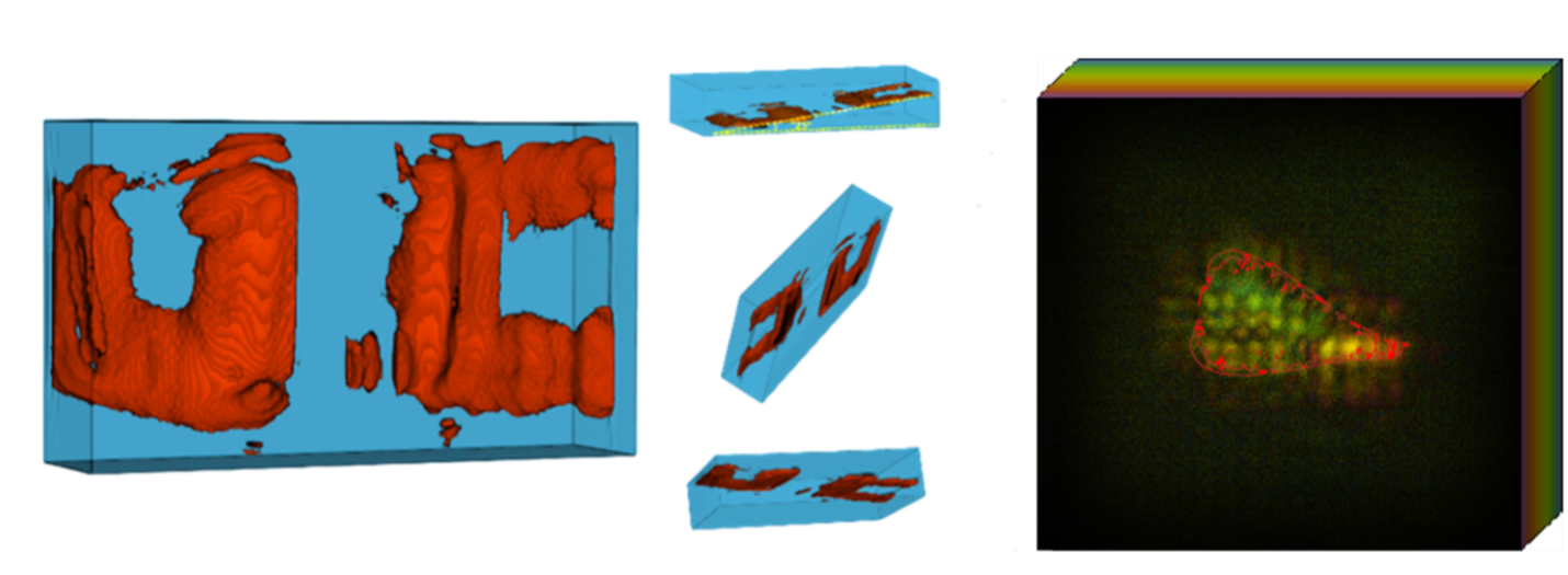
3D Imaging with XACT
The above graphics illustrate what 3D imaging with XACT looks like with examples of the UC logo on the left and a bone sample on the right.

Challenges and Future Directions
While the potential of XACT is immense, current limitations include resolution constraints tied to the frequency and size of the ultrasound detectors. Future improvements, such as higher-frequency transducers and advanced reconstruction algorithms powered by deep learning, could further enhance its performance.

Redefining Imaging Across Fields
The ability to achieve 3D imaging from a single X-ray projection positions XACT as a transformative tool not only for medical diagnostics but also for nondestructive testing in engineering and material science. Its innovative approach eliminates the need for rotational access, opening new possibilities for imaging in constrained environments.

XACT also represents a leap forward in imaging technology, combining reduced radiation exposure, compact system design and unprecedented efficiency. As this technology continues to evolve, it has the potential to redefine medical and industrial imaging, bringing us closer to a future where high-resolution, low-dose 3D imaging is the norm in healthcare and beyond.

Filed Under: News

Protoacoustic Imaging for Precision Proton Therapy Recognized as Cancer Breakthrough at ASTRO 2024

October 26, 2024 by liangzhx

Thanks American Association of Physicists in Medicine’s Science Council Chair Dr. Jan Seuntjens for highlighting our work on protoacoustic imaging for precision proton therapy on the great American Society for Radiation Oncology (ASTRO 2024) as the Cancer Breakthrough this year! It is a great collaborative effort with Dr. Yong Chen’s group and Dr. Lei Ren ‘s group. Thanks for the hard work from all my students and fellows, I am so proud of you all!!

Filed Under: News

Dr. Shawn Xiang Presents Groundbreaking Advances in Radiacoustic Imaging at NCI Webinar

October 26, 2024 by liangzhx

Dr. Shawn Xiang delivered a webinar for the National Cancer Institute (NCI) on October 7, 2024, titled “Theranostics with Radiation-induced Ultrasound Emission (TRUE).” In this presentation, Dr. Xiang discussed three groundbreaking projects related to radiacoustic imaging:

  1. In vivo dosimetry and image-guided precision radiotherapy
  2. Precision Nano-Knife
  3. XACT: A portable and ultrafast CT

For more details, please visit the NCI website: NCI Imaging Webinar.

Filed Under: News

Dr. Xiang Featured in AAPM Interview and Research Spotlight

August 16, 2024 by liangzhx

Dr. Shawn Xiang was interviewed by Dr. Rongxiao Zhang and Richard Dargon from AAPM, where he shared his insights on image-guided FLASH radiotherapy. During the interview, Dr. Xiang introduced the concept of radiation-induced acoustic imaging, a groundbreaking technology developed by the TRUE lab, which plays a crucial role in guiding FLASH RT with greater precision. He was joined by Dr. Brian Pogue, the department chair of Medical Physics at the University of Wisconsin-Madison, who discussed his work on Cerenkov imaging and other innovative technologies that are advancing the clinical application of FLASH RT. For more details, you can read the full interview here: AAPM Interview.

Filed Under: News

Congrats to Lauren Tidyman for Winning the UCI UROP Summer Research Award!

August 2, 2024 by liangzhx

Lauren Tidyman just finished her 1st undergraduate study at UCI, and she is working with PhD student Yifei Xu on electroacoustic tomography research. Congrats Lauren for winning the UCI UROP summer research award!

Filed Under: News

Former PhD student Siqi Wang’s work won the Science Council innovation physics award at AAPM annual conference!

August 2, 2024 by liangzhx

Thanks AAPM 2024 for giving us the Science Council innovation physics award !
The credit goes to my former PhD student Siqi Wang and Gilberto Gonzalez with Dr. Yong Chen at OUHSC! This work on protoacoustic imaging for localizing the Bragg peak has made the impact in the medical physics community. Siqi is now a postdoc scholar at Stanford University. Congrats Siqi for your wonderful work at TRUE lab!

Filed Under: News

PhD student Kristina Bjegovic got the Best in Physics Award from AAPM annual meeting!

August 2, 2024 by liangzhx

At this year’s AAPM annual meeting in Los Angeles, our PhD student Kristina Bjegovic won the prestigious Best in Physics Award for her work on “Protoacoustic Monitoring of GRID FLASH Proton Therapy.” This is a highly competitive award with only a few recipients. Thanks to the collaborative efforts with the clinical team at OUHSC and Dr. Yong Chen’s team!

Filed Under: News

Dr. Prabodh Pandey Publishes Research on Protoacoustic Microscopy

February 29, 2024 by liangzhx

TRUE Lab Postdoctoral Researcher Dr. Prabodh Pandey Advances Photoacoustic Microscopy with Innovative Approach. He introduces the concept of employing a low-energy proton beam to generate ultrasound signals for microscopic imaging. This approach, utilizing a finely focused proton beam, promises nanometer-level resolution combined with ultrasound’s ability to enhance penetration depth, enabling super high-resolution imaging of living cells.

Filed Under: News

TRUE lab undergraduates won UROP fellowships

February 19, 2024 by liangzhx

Three undergraduate research assistants from the TRUE lab, Shreyas Srinivasan, Yuxuan Bai, and Sanjana Khedikar, have been awarded UROP fellowships for their projects. Congratulations!

Filed Under: News

Ph.D student, Yifei Xu’s Research Paper Featured in Editors’ Choice in year 2023

January 15, 2024 by liangzhx

Yifei’s Study on Electroacoustic Tomography for Real-Time Monitoring of Electroporation Recognized on Nature.com: https://www.nature.com/articles/s44172-023-00151-7. This is a selection of short Research Highlights describing some of the favourite Communications Engineering publications of 2023 by the Editorial Board and Editorial Team. Congrats!

Filed Under: News

  • Page 1
  • Page 2
  • Page 3
  • …
  • Page 5
  • Next Page »

Recent Posts

  • UC Irvine researchers reveal a revolutionary new approach for 3D CT imaging from a single X-ray projection
  • Protoacoustic Imaging for Precision Proton Therapy Recognized as Cancer Breakthrough at ASTRO 2024
  • Dr. Shawn Xiang Presents Groundbreaking Advances in Radiacoustic Imaging at NCI Webinar
  • Dr. Xiang Featured in AAPM Interview and Research Spotlight
  • Congrats to Lauren Tidyman for Winning the UCI UROP Summer Research Award!

© 2025 UC Regents